
Why the Protocol Matters More Than the Material
A practitioner can use premium ceramic materials and still deliver a failing restoration if the adhesive protocol is not correctly executed. Conversely, even modestly priced composite materials, bonded with the full biomimetic protocol, outperform expensive ceramics placed over incorrectly conditioned dentin in long-term clinical outcomes.
The biomimetic bonding protocol was developed from foundational research by Pascal Magne at USC and refined into clinical teaching by David Alleman and Jon Deliperi through the Six Lessons curriculum. It treats the adhesive interface not as a means of "sticking things together" but as an engineered structural zone that must replicate the biomechanical properties of the enamel-dentin junction it is replacing.
Pillar 1 — Selective Enamel Etching
The first decision in the biomimetic protocol is where to apply phosphoric acid etchant. Conventional total-etch protocols apply 37% phosphoric acid to both enamel and dentin simultaneously for 15–30 seconds. This maximizes enamel bond strength but simultaneously over-etches dentin, dissolving the smear layer and collapsing the collagen fibril network — reducing the quality of the subsequent dentin bond.
The biomimetic approach is selective: phosphoric acid is applied to enamel margins only, for full etch duration. Dentin is conditioned with a self-etch adhesive primer in separate steps — its lower acidity conditions the smear layer without destroying the collagen architecture. This produces high enamel bond strength (critical for the peripheral seal zone) while preserving dentin collagen integrity for maximum hybrid layer formation.
Pillar 2 — Immediate Dentin Sealing (IDS)
IDS is applied immediately after preparation and enamel etching — before any impression, scan, or provisional placement. The hydrophilic bonding resin penetrates freshly cut dentin tubules at maximum receptivity (freshly cut, fully hydrated, physiologically active dentin achieves the highest adhesive bond strength). Light-curing creates a sealed, cross-linked resin layer that:
- Blocks bacterial and salivary contamination of dentin tubules during the provisional phase.
- Prevents desiccation and collagen collapse that degrades bond strength over time.
- Creates a pre-polymerized surface that, when reactivated at cementation, produces 80–120% higher bond strength than deferred bonding (bonding only at cementation).
After IDS light-curing, a thin layer of glycerin is applied and re-cured to eliminate the oxygen-inhibited surface layer that would otherwise contaminate provisional cement and prevent reactivation. See the full IDS guide for detailed technique.
Pillar 3 — C-Factor Management
C-factor (configuration factor) is the ratio of bonded composite surfaces to free (unbonded) composite surfaces in a given increment. As C-factor increases, polymerization shrinkage stress has fewer free surfaces to flow toward — concentrating stress at the bonded interfaces and risking adhesive failure or cuspal deflection. High C-factor configurations (deep Class I cavities, box-shaped preparations) are the primary cause of post-operative sensitivity and marginal leakage in direct composite restorations.
Biomimetic protocol manages C-factor by:
- Using horizontal increments no thicker than 1.5–2 mm, each with at least one free surface.
- Placing the first increment as a thin "liner" over the deepest, highest C-factor zone — using low-modulus, low-shrinkage flowable composite rather than a high-filler packable composite.
- Strategically leaving one wall free during each increment placement, then covering it with the subsequent increment after the previous one has cured and its shrinkage vector has been absorbed.
- Using low-shrinkage, high-filler composite formulations (shrinkage <2.0%) for the bulk of the preparation base.
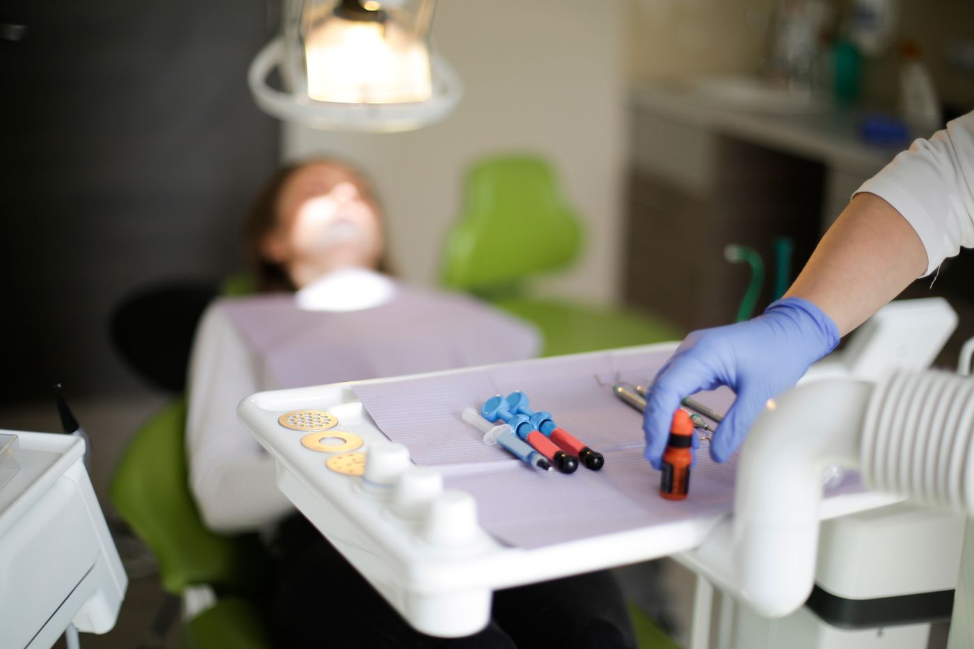
Pillar 4 — Stress-Reduced Composite Layering
The stress-reduced composite base serves two structural roles:
- Biomechanical cushion: Composite has a modulus of elasticity (15–20 GPa) intermediate between dentin (15–20 GPa) and ceramic (70–100 GPa). Placing a composite layer between dentin and ceramic creates a modulus gradient that reduces peak interfacial stress concentration at the ceramic-tooth junction under functional load — mimicking the enamel-over-dentin structural gradient of the natural tooth.
- Polymerization shrinkage absorption: By placing composite in stress-controlled increments before the ceramic restoration is bonded, all polymerization shrinkage stress is absorbed and dissipated before the ceramic restoration adds its own volume. The ceramic-to-composite bond at cementation is therefore formed on a dimensionally stable, already-polymerized substrate — not on a composite that is still trying to shrink.
Pillar 5 — Peripheral Seal Zone Creation
The peripheral seal zone is the band of enamel at the preparation margins, selectively etched with phosphoric acid and bonded with the highest achievable enamel bond strength (25–40 MPa). This enamel seal band acts as the outer perimeter of the adhesive system — the last line of defense against marginal microleakage.
Preparation design in biomimetic dentistry specifically preserves as much marginal enamel as possible, even when cavity extension would be geometrically easier with dentin margins. Internal dentin bonding can be excellent, but enamel bonding is more durable, more predictable, and less susceptible to hydrolytic degradation over time. A restoration whose margins are entirely in enamel — all other things being equal — will outlast a restoration whose margins are in dentin.
The Protocol at Cementation: Reactivation and Assembly
At the cementation appointment, the IDS surface is lightly air-abraded with 27-micron alumina to remove provisional cement residue and expose fresh adhesive surface. The ceramic intaglio is treated with hydrofluoric acid etch and silane coupling agent. Bonding agent is applied to both the reactivated IDS surface and the silanized ceramic. Dual-cure adhesive resin cement is applied to the ceramic, the restoration is fully seated, excess cement is removed in the gel phase, and final light-curing is performed through the ceramic (dual-cure is required for adequate depth of cure in opaque ceramic restorations where light transmission is limited).
The Diagnostic Question for Any Indirect Restoration
Before any indirect restoration, ask your dentist to describe what they do to the tooth between preparation and cementation — specifically with regard to the freshly cut dentin. If the answer is "take the impression and place a temporary," you are not receiving the biomimetic protocol. If the answer describes a multi-step adhesive sealing process at the preparation appointment, you are.
Frequently Asked Questions
Does the biomimetic protocol take longer than conventional?
Yes — approximately 15–25 minutes longer at the preparation appointment, due to IDS, selective etching, and composite basing. The cementation appointment is similar in duration. The additional chairside time is offset by dramatically lower rates of post-operative sensitivity, remakes, and long-term restoration failures.
Can any dentist learn this protocol?
Yes — the biomimetic protocol is a set of techniques, not restricted to any specialty. It requires dedicated post-doctoral training through programs like the Alleman Center Six Lessons curriculum or AOBMD fellowship preparation. It is not taught comprehensively in most dental schools, which is why credential verification matters when seeking biomimetic care.
Is the biomimetic protocol only for posterior teeth?
No — while the protocol was developed primarily for posterior indirect restorations, the principles of IDS, selective enamel etching, and C-factor management apply to anterior veneers, direct composites, and any adhesive restoration where dentin is exposed during preparation.